Radiology
Expert Imaging With a Personal Approach
Radiology
Make an Appointment
We have locations throughout Manhattan and Westchester. View all locations and contact info.
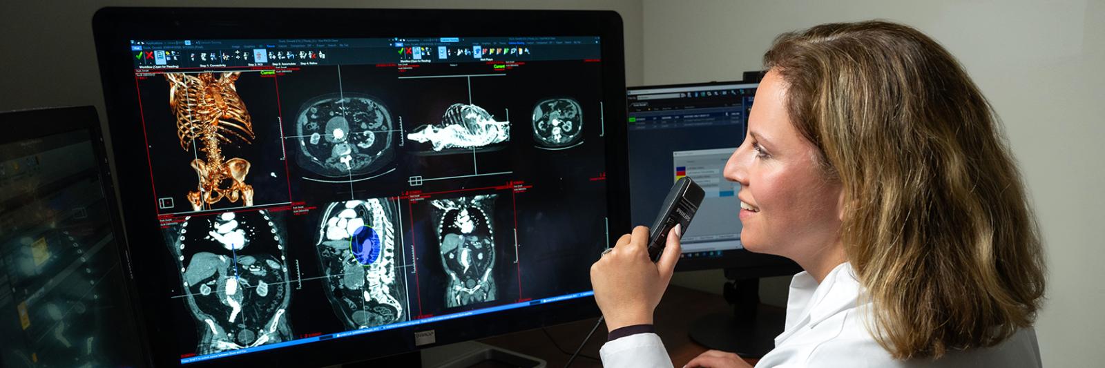
Radiologists play a critical role in the early detection, diagnosis, and even in the treatment of disease. Our radiologists are nationally-recognized experts who have a deep understanding of disease, often catching important details that may otherwise be overlooked.
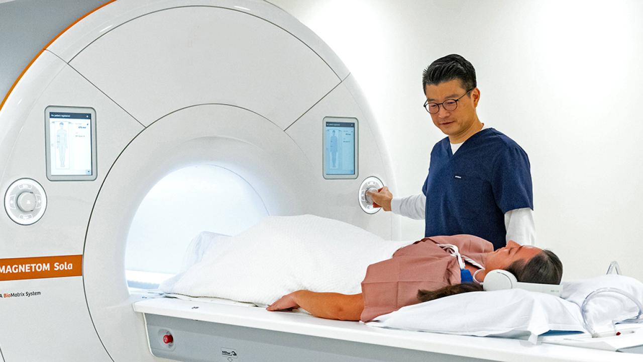
When you choose Columbia Radiology, you're choosing to be affiliated with NewYork-Presbyterian Hospital, one of the top rated hospitals in the country. You'll have access to the most current technology and image-guided therapies along with radiologists and technicians who perform and interpret more than 800,000 procedures and scans each year. Our commitment to expert, personal care means that your radiologist will partner with your physician and become an integral part of your healthcare team.
Radiology Services


The Department of Radiology

The Department of Radiology at Columbia's Vagelos College of Physicians & Surgeons is committed to the pursuit of scientific excellence in imaging research and developing the next generation of radiologists and imaging scientists.